
Reproduction is the formation of new individuals on the pattern of existing ones. Man, like most other large, advanced organisms, propagates himself by sexual reproduction, a process involving a parent of either sex and resulting in variable offspring. Each parent provides special reproductive cells, or gametes-females produce egg cells or ova, and males produce spermatozoa – which cannot live on their own. Brought together in the female reproductive tract and allowed to fuse, the two form a single, fertile zygote, which grows into a new individual. The new organism resembles both of its parents in important issues, but differs slightly from them in detail; some of its characteristics may be copied faithfully from one or other parent, some may be a blend of parental characteristics; and some may be inherited from grandparents or more remote kin. The combination of different genes, which are the determining factors of all our characteristics, produces new individuals, which are unique.
Irrespective of race, colour or creed, all human beings are divided into two groups- male and female. The most important single difference between the two is the structure and housing of the gonads (ovaries and testes) – the organs that from and nurture the reproductive cells- and the arrangement of internal and external accessory organs, the genitalia. The sexes also differ in body size and proportion, skeletal structure, the distribution of body hair, metabolic rate, temperament, and in many other ways. Some differences are entirely physical, dictated by the body itself. Others are largely or entirely cultural, imposed by the society we live in.
The sex of an infant is determined at its conception by the pattern of chromosomes contained in the newly formed zygote. Every normal human cell (other than a gamete) contains in its nucleus 46 chromosomes, in dividing cells these can often be seen arranged in 23 matching pairs. Forty-four of the chromosomes are autosomes, not directly concerned with sex determination. The remaining two are sex chromosomes, which from an identical pair in females (XX). But an ill matched pair in males, one being shorter than the other (XY). The short Y chromosome is assumed to carry the male determining factor.
When gametes are being formed in the gonads, the specialized cells, which produce them, split into two, one half of each chromosome pair passing to each gamete. Ova formed in this way necessarily contain22 autosomes and one X chromosome. Spermatozoa, on the other hand, contain 22 autosomes and either an X or a Y chromosome, because they are derived from male XY-endowed body cells. Thus there is only one kind of ovum, but two kinds of spermatozoa. An ovum fertilized by an X-chromosome-carrying sperm will become an XX zygote and develop into a female embryo. One fertilized by a Y-chromosome-carrying spermatozoon becomes an XY zygote, which develops into a male embryo.
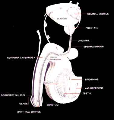
Spermatozoa, produced in the testes, travel throughout the vas deferens, bypass the bladder and enter the ejaculatory duct, a muscular-walled tube, here, alkaline secretions produced by tow seminal vesicles (which supply solutions of fructose and ascorbic) and the prostate gland (which adds cholesterol, fatty acids, phospholipids and other nutrients), surround the spermatozoa, forming the grayish-white fluid known as semen, which transports spermatozoa out of the male body. Semen is an alkaline substance because spermatozoa survive longer in an alkali than in acid. The semen passes into the urethra and is forced out through its opening during ejaculation. In a male organism, about three to five milliliters of semen are ejaculated. Each milliliter may contain more than 60 million spermatozoa, each capable of fertilizing an egg inside a woman’s body
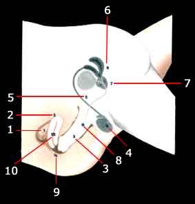
Seen here in section are the male and female gentalia during sexual intercourse.From the overy
1)an ovum released at the previous time of ovulation is propelled along the Fallopian tube towards the uterus
2)The penis
3)is the means by which spermatozoa,formed in the testis
4)and passed along the vas deferens
5)will be deposited in the female tract during orgasm.The seminal vesicles
6)and the prostate gland
7)add their own secretions to the seminal fluid,which is pumped through the ejaculatory duct
8)by waves of muscular contraction, and forms a pool near to the cervix of the uterus
9)The uterus,which during intercourse has moved up and away from the upper end of the vagina,now descends,so that the cervix dips into the pool of semen. Spermatozoa awim through the uterine cavity
10)Fartilization usaually takes place in one of the Fallopian tubes.
In the shaft of the penis, the urethra, through which urine or spermatozoa pass out of the body, is surrounded by the corpora cavernous, tough-walled sacs containing spongy tissue, which fill with blood under pressure during erection. The penis is covered with skin, which forms a loose fitting shroud over the sensitive glans penis. A man has two testes, oval organs about 39 millimeters long by 27 millimeters wide, carried in a pouch of skin called the scrotum. Each testis is divided into lobules containing long, narrow coiled somniferous tubules. Cells lining the somniferous tubules form spermatozoa, releasing millions for temporary storage in the epididymis. When a man is sexually aroused, the spermatozoa pass into the vas deferens and on into the urethra.
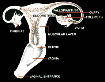
The gonads are the organs, which form and nurture the reproductive cells. In a woman, they take the form of ovaries, which produce the ovum or egg. The male gonads are the testes, which produce the spermatozoa. Fundamentally, the two systems are similar. In both sexes the two gonads are separate organs carrying out the same function. Both ovaries and testes are linked to the external reproductive organs- the vagina in the female and the penis in the male- by a system of ducts and sinuses. In the female, the individual ducts join to form the uterine cavity. In the male, they join at the beginning of the urethra, making one channel to the outside world. In the embryo, two pairs tubes, the Mullerian and the Wolffian ducts, link the gonads to the area which form the External reproductive organs. About two months after conception, the foetus assumes its sexual identity. If female, the gonads become ovaries, the Wolffian ducts degenerate and the Mullerian ducts develop into the Fallopian tubes, uterus and vagina. If the foetus is male, the Mullerian ducts disappear, the Wolffian ducts develop into the seminiferous tubules and spermatozoa – carrying ducts, and the gonads develop into testes. At puberty, hormones released by the anterior pituitary to produce their reproductive cells and to secrete hormones of their own activate the gonads.
Both male and female gonads serve a dual purpose, producing both reproductive cells and sex hormones. In the male, the testes secrete testosterone, the hormone responsible for the development of male characteristics, stimulating the growth of skeletal muscle, the production and distribution of body and facial hair, the deepening of the voice and the rapid development of both internal and external sexual organs. It is secreted in response to another hormone, the interstitial-cell-stimulating hormone (ICSH), which is secreted by the anterior pituitary. In the female, the ovaries secrete two hormones, estrogen and progesterone. In the first week of the menstrual cycle, the pituitary secretes follicle-stimulating hormone (FSH).
Mature follicles secrete estrogen, which is responsible for the development of female characteristics – the growth of the breasts, the distribution of fat and of body hair and also stimulate the growth of the uterine lining. When the follicle is about to burst, the estrogen level drops, causing the pituitary to secrete the luteinizing hormone (LH), triggering ovulation. The ruptured follicle becomes the corpus lutetium, secreting progesterone, which prepares the lining of the uterus for the reception of a fertilized egg. If none appears, the level of both estrogen and progesterone falls, beginning the cycle again.
The ovaries are situated within the basin formed by the pelvic bones. At birth, each ovary contains several hundred thousand minute sacs called follicles. Only between four and five hundred of these develop to maturity. When they are ready to burst and shed their ovum. Unlike the male testis, which produces millions of spermatozoa, the ovaries usually release one ovum about every four weeks; the ovum may come from either ovary. This process begins just after puberty and continues for about 30 years. Each time an ovum is released, it is picked up a frond-like fimbria and taken into Fallopian or uterine tube, where fertilization normally occurs. The ovum is wafted along by the cilia lining of the uterus, the enbonmetrium, and a mucous membrane specifically designed to receive and retain it, and richly supplied with blood to feed a developing foetus. If the ovum of the month is not fertilized, this lining breaks up and is washed away, mixed with blood form the vessels to which it was attached, through the cervix and vagina. This monthly flow of blood is known as menstruation. The vagina is a highly sensitive muscular tube lined with mucous membrane, which receives the penis during intercourse and conducts the ejaculated spermatozoa to the cervix, through which they pass into the uterus and up to the fallopian tubes.
It is therefore the father’s contribution to the zygote that determines the sex of an infant and in theory, equal numbers of each sex should be produced, in fact, Y-chromosome-carrying spermatozoa are very slightly smaller than X-carriers, and faster moving, and seem able to fertilize more than their fair share of Ova. At the moment, boys outnumber girls at birth by about 21:20.
The reproductive organs appear early in the growing embryo as tiny knots of tissue close to the spine on the abdominal wall. Two pairs of tubes, the Mullerian and the Wolffian ducts, link the developing gonads with a small hollow- the urogenital sinus-at the rear end of the embryo. Up to the eighth week after conception, there is little to distinguish male from female. The presence or absence of the Y chromosome makes itself apparent later. In the female embryo, where both chromosomes are X-carriers, the developing gonads become ovaries, the Wolffian ducts disappear, the Mullerian ducts differentiate into the female reproductive tract-paired Fallopian tubes, uterus and vagina and from the urogenital sinus develop the paired labia majora and labia minora and associated glands of the female. In the presence of both X and Y chromosomes in the male embryo, the gonads become testes, the Wolffian ducts become sperm-carrying ducts and tubules, and the Mullerian ducts degenerate. The urogenital sinus becomes part of the urethra and the tissues on either side join along the midline to form the sac of the scrotum. This remains empty until the seventh or eighth month of foetal life, when the testes descend into it form the abdominal wall. The phallus is initially a small bump on the front edge of the urogenital sinus. In the male, it grows larger, acquiring two longitudinal masses of spongy, erectile tissue and the tube of the urethra, from the penis-the erectile organ through which spermatozoa are transmitted from the male into the female reproductive tract. In the female, it is small, forming the sensitive and erectile clitoris.
Throughout early life, the reproductive organs remain small and non-functional, a source of mild interest and pleasant sensation to their owners. Shortly before puberty, gonadotrophic hormones produced in the anterior lobe of the pituitary gland cause the ovaries and testes to develop and secrete hormones of their own. These in turn stimulate the growth of the genitalia, and the changes of body form which are associated with adolescence. At the same time, the gonads start to produce their gametes, at first sporadically and then continuously. Girls begin to ovulate, settling eventually into a regular cycle in which a single ovum is released, and the reproductive tract made ready for pregnancy once every 28 days. In boys, millions of spermatozoa begin to form in the somniferous tubules of the testes and the seminal vesicles and prostate secrete a nutritious liquid medium for them.
Not surprisingly, young people find these changes of body structure interesting and exciting and very sensibly try out for themselves the range of new sensations associated with them. Most boys and many girls, discover sexual excitement and orgasm through masturbation at this stage, learning to relieve their nervous tensions in a natural way.
Choose color style